LINEBook via LINE
Call LINE Book
Abnormal Circulating Cell Screening

ตรวจเซลล์ผิดปกติในเลือด (CEC)

Abnormal Circulating Cell Screening (CEC)

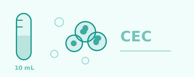
A single 10 mL blood draw, looking for a cell that should not be there in someone healthy. Results in 72 hours. A strong signal to go for a wider vascular workup.
Available Packages
Abnormal Circulating Cell Screening (CEC)
Early Signal
Abnormal Circulating Cell Screening (CEC)
· 1 tests
The cell screen on its own, from a single 10 mL blood draw, read by multiplex cytopathology.
Tests Included
  • Clustered abnormal endothelial cells (CEC) by multiplex cytopathology
  • A written report explained in both Thai and English
  • A follow-up pathway for your physician if the result is abnormal
Who It's For
Anyone who wants the cell screen alone, or who has had tumour markers done recently.
Best
Abnormal Circulating Cell Screening (CEC) + Tumour Marker Panel
Early Signal
Abnormal Circulating Cell Screening (CEC) + Tumour Marker Panel
· 11 tests
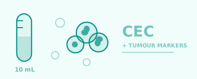
The cell screen combined with a tumour marker panel covering both men and women, from a single 10 mL blood draw.
Tests Included
  • Clustered abnormal endothelial cells (CEC) by multiplex cytopathology
  • A tumour marker panel covering the major organ groups, for both men and women
  • A written report explained in both Thai and English
  • A follow-up pathway for your physician if the result is abnormal
Who It's For
Anyone wanting an early signal before symptoms appear, or with a family history that worries them.

Want the tumour markers alone at a lower price? See the tumour marker panels.

ISO 15189 laboratory Accredited by the Department of Medical Science CE-IVDR certified 10 mL blood · results in 72 hours
What the pathologist sees
Single endothelial cells among red blood cells
Normal blood Single endothelial cells circulate at low levels in everyone
A cluster of abnormal, irregularly nucleated cells
What this test looks for Clustered, aneuploid and abnormally nucleated — not found in healthy people

Schematic illustration, not a microscope image — the same principle as a Pap smear, reading the shape of a cell. What differs is the sample it is read from: blood.

What the test looks for
Cells shed from a vessel wall
Where a blood vessel is damaged, the endothelial cells lining it detach into the bloodstream. This test collects them from 10 mL of blood and reads them under a microscope.
The abnormality is the clustering, not the presence
Ordinary single endothelial cells circulate at low levels in everyone. What is not found in healthy people is cells that are clustered, aneuploid, and abnormally nucleated.
We read the cell, not a number
The same principle as the Pap smear, which has used cell shape to flag disease for over seventy years. What differs is the sample it is read from: blood.
From a 10 mL draw to a result
  1. 1 Heparin blood A single 10 mL draw
  2. 2 Red cell lysis Healthy red cells removed
  3. 3 White cell depletion Our own separation method
  4. 4 Multiplex staining Several markers at once
  5. 5 Cytoscreening Shape and staining, read together

Separating a handful of rare cells from several billion blood cells is one problem; reading the shape and the staining pattern of that same cell is another. X-ZELL developed both. Every sample is processed in Bangkok at an ISO 15189 laboratory accredited by the Department of Medical Science — the only CE-IVDR certified multiplex cytopathology method available, and the only accredited CEC test in Thailand.

Why cells and tumour markers together

Blood tumour markers on their own are highly non-specific. A raised value can come from inflammation, infection, or something entirely benign. That is why they should not be used to decide whether a particular disease is present — and why they work as a prompt to look further.

The same is true of an abnormal cell finding. It says something in the body is worth a closer look. It does not say what.

Both are non-specific signals, but they are different kinds of signal — one read from a cell, one measured from a protein. When the two point the same way, your doctor starts from firmer ground than either would give alone. And both lead to the same next step: targeted follow-up rather than a blind search.

The scientific evidence

Abnormal circulating endothelial cells are not something X-ZELL found on its own. Independent groups in several countries have published on them for decades, and that literature falls into three groups.

Vascular disease
Work going back to 1997 found these cells raised in acute myocardial infarction, vasculitis and autoimmune disease. This literature developed entirely separately from the cancer one.
Cancer
Reported across more than 13 cancer types — prostate, breast, colorectal, lung, ovarian, liver, pancreatic and others — by unrelated groups in multiple countries.
Healthy people
Separate studies exist for the sole purpose of establishing what a normal level looks like. That is what makes it possible to say what counts as abnormal.
Read the source research

A few of the anchor papers in each group, linked straight to the abstract. X-ZELL's own study is one entry on this list, not the list itself.

What your result means
If your result is normal
No clustered abnormal cells were found. Carry on with your regular check-ups. This is a good sign, not a guarantee, and your doctor can explain what it does and does not rule out.
If your result is abnormal
An irregularity was found. That is not a diagnosis, and not a reason to panic — often the cause is very treatable. Your physician will review your health history and suggest targeted follow-up.
What happens after an abnormal result
1
Rule out temporary causes
History alone excludes most of them: smoking in the last 24 hours, a recent infection or acute illness, major trauma or surgery, and any known blood disorder.
2
Vascular workup
The most likely finding in a wellness population: lipids, hs-CRP, blood pressure, kidney function and HbA1c. See Cardiovascular & TMAO testing.
3
Wider workup if still unexplained
Where the first two steps do not account for the result: autoimmune screening, age- and risk-appropriate screening, organ-specific imaging where indicated, and a repeat test to confirm the finding persists.

This is guidance, not a fixed protocol. The workup remains the treating physician's clinical judgement.

FAQ
Can this test tell me whether I have cancer?

No. This is not a diagnostic test for any single disease. An abnormal result says a cell was found that should not be there in someone healthy — which can have several causes, including vascular disease and autoimmune conditions. Your doctor weighs it against your health history to decide what to check next.

How is this different from ordinary tumour markers?

Tumour markers measure protein levels in blood. This test reads the shape and staining of actual cells. Neither is specific on its own, which is why this package includes both — they are different kinds of signal and they inform each other.

How should I prepare?

Avoid smoking for at least 24 hours before the draw — it interferes with the result. If you have recently had an infection, an acute illness, a major injury or surgery, tell our team in advance so we can schedule at a more suitable time.

When do I get the result?

Within 72 hours of the blood draw, delivered by e-mail or LINE with an explanation of what it means.

How often should I repeat it?

Typically every 6 to 12 months, depending on your previous result and your own risk profile. After an abnormal result your doctor may suggest repeating it sooner to confirm the finding persists.

Open Mon–Fri 09:00–17:00

Private service · Results via E-mail/LINE · Expert team